Anatomy, Radiographic Anatomy and Cephalometric Landmarks of Craniofacial Skeleton, Soft Tissue Profile.
A lateral cephalogram is one of the orthodontic records that provides information about the sagittal and vertical relations of:
• the craniofacial skeleton;
• the soft tissue profile;
• the dentition;
• the pharynx; and
• the cervical vertebrae.
These structures and their relationships to each other are scrutinized by means of linear and angular measurements as well as by the use of ratios based on the various cephalometric landmarks. These cephalometric landmarks should be identified; fight that began at the errors in their identification can be minimized by a thorough knowledge of the anatomy of the skull and by an awareness of the close correspondence between gross anatomy and radiographic appearance of each structure and the detailed criteria for identification of each anatomical cephalometric point.
CRANIOFACIAL SKELETON
FRONTAL BONE
Anatomy

Photograph of the lateral aspect (A) and the medial aspect (B) o f the frontal bone.
1 frontal bone 8 zygomatic bone
2 parietal bone 9 frontonasal suture
3 coronal suture 10 frontomaxillary suture
4 sphenoid bone 11 frontozygomatic suture
5 ethmoid bone 12 frontal sinus
6 nasal bone
7 maxilla
The frontal hone (1) forms the anterior part of the cranial vault. It joins posteriorly with the parietal bones (2) at the coronal suture (3). It joins inferiorly with the sphenoid bone (4) and the ethmoid bone (5) at the frontosphenoethmoidal suture. Anteriorly, it joins with the nasal bones (6), with the maxilla (7), and the zygomatic bone (8) at the fron-tonasal suture (9), the frontomaxillary suture (10), and the frontozygomatic suture (11), respectively. The lower anterior part of the frontal bone forms the roof of the orbit, and laterally its zygomatic process joins with the frontal process of the zygomatic bone forming the lateral border of the orbit. The frontal sinus (12) lies in the frontal bone, in an area superior to the articulation with the nasal bone.
Radiographic anatomy

Radiograph of the lateral view of the frontal bone.
2 external cortical plate of frontal bone
3 internal cortical plate of frontal bone
4 frontal sinus
5 frontonasal suture
6 nasal bone
7 frontosphenoethmoidal suture
8 endocranial surface of frontal bone
9 exocranial surface of frontal bone
10 lesser wing oi sphenoid bone
11 anterior clinoid
12 anterior margin of zygomatic process of frontal bone
13 posterior margin of zygomatic process of frontal bone
14 anterior margin of frontal process of zygomatic bone
15 posterior margin of frontal process of zygomatic bone
Starting from the upper anterior part of the skull at the coronal suture (1), the frontal bone appears as two radio-opaque lines that descend parallel to each other. The outer radio-opaque line represents the external cortical plate of the frontal bone (2), and the inner line represents the internal cortical plate (3), which forms the anterior border of the anterior cranial fossa. These two parallel lines diverge at the forehead area where the frontal sinus(4) appears as a radiolucent area between them. The external cortical plate terminates at the anterior part of the frontonasal suture (5), which appears as a radiolucent line between the frontal and the nasal bones (6). The internal cortical plate extends horizontally and posteriorly, thus terminating at the small radio-opaque triangular area that represents the frontosphenoethmoidal suture (7),
Above the horizontal part of the internal cortical plate there are two radio-opaque lines. The uppermost of these two lines, which appears as a wavy radio-opaque line, represents the endocranial surface of the frontal bone (8), which forms the floor of the anterior cranial fossa. The harmonious radio-opaque curve below the wavy line represents the exocranial surface of the frontal bone (9), which forms the roof of the orbit. This line extends posteriorly to the lesser wings of the sphenoid bone (10) and to the anterior clinoid (11). Anteriorly, it starts at the area of the frontal sinus where the junction of the roof of the orbit and its lateral border can be identified as an angular radio-opaque shadow. The lateral border of the orbit appears as a curved radio-opaque line, which represents the anterior margin of the zygomatic process of the frontal bone (12). At the same area, the posterior margin of the zygomatic process of the frontal bone (13) can be identified as a radio-opaque line descending parallel behind the lateral border of the orbit. These two lines merge with the radio-opaque lines of the anterior and posterior margins of the frontal process of the zygomatic bone (14, 15).
Cephalometric landmarks
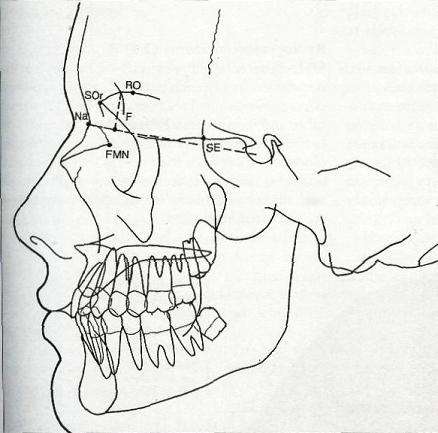
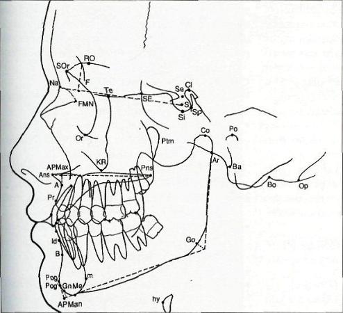
F-point F (constructed) - this point approximates the foramen caecum and represents the anatomic anterior limit of the cranial base, constructed as the point of intersection of a line perpendicular to the S-N plane from the point of crossing of the images of the orbital roofs and the internal plate of the frontal bone (Coben);
FMN - frontomaxillary nasal suture - the most superior point of the suture, where the maxilla articulates with the frontal and nasal bones (unilateral); FMN is on the anterior cranial base, unlike Na , and can therefore also be used for measuring or defining the cranial base (Moyers, 1988);
Na - nasion - the most anterior point of the fron-tonasal suture in the median plane (unilateral);
SE - sphenoethmoidal - the intersection of the shadows of the greater wing of the sphenoid and the cranial floor as seen in the lateral cephalogram;
SOr - supraorbitale - the most anterior point of the intersection of the shadow of the roof of the orbit and its lateral contour (bilateral) (Sassouni);
RO - roof of orbit - this marks the uppermost point on the roof of the orbit (bilateral) (Sassouni).
SPHENOID BONE
Anatomy

Photograph of the lateral aspect (A) and the medial aspect (B) of the sphenoid bone.
1 maxilla
2 palatine bone
3 ethmoid bone
4 frontal bone
5 lesser wing of sphenoid bone
6 greater wing of sphenoid bone
7 pterygoid process of sphenoid bone ,
8 sphenoid sinus
9 sella turcica
10 anterior clinoid
11 posterior clinoid
12 dorsum sellae
13 optic canal
14 parietal bone
15 medial pterygoid plate
16 lateral pterygoid plate
17 pterygoid hamulus
18 pterygomaxillary fissure
19 sphenopalatine foramen
Anteriorly, the sphenoid bone articulates with the maxilla (1) and the palatine bone (2); anterosuperiorly it articulates with the ethmoid bone (3) and the frontal bone (4) at the frontosphenoethmoidal suture. It consists of the body and the three paired processes - the lesser wings (5), the greater wings (6) and the pterygoid process (7).
The sphenoid body is occupied by the two air-filled cavities called the sphenoid sinus (8). Its superior surface has a deep depression of a saddlelike appearance called the sella turcica (9), which houses the pituitary gland. The anterior limit of the sella turcica is the anterior clinoid (10), the posterior limit is the posterior clinoid (11) and the dorsum sellae (12).
The lesser wings of the sphenoid (5) project anteriorly to the sella turcica (9), where the optic canals (13) can be seen. The superior surfaces of the lesser wings form the floor of the anterior cranial fossa and their inferior surfaces form the most posterior part of the orbital roof.
The greater wings (6) project from the postero-lateral portion of the body. They articulate laterally with the frontal (4) and parietal (14) bones, and posteriorly with the squamous portion of the temporal bone.
The pterygoid processes (7) project inferiorly from the root of the greater wings (6). Each process consists of two plates, the medial and the lateral pterygoid plates (15,16), which are separated by the deep fossa. The inferior end of the medial pterygoid plate is a thin curved process called the pterygoid hamulus (17).
Between the posterior border of the maxilla (1) and the anterior surface of the pterygoid process (7) is the pterygomaxillary fissure (18), with an inverted teardrop shape. The sphenopalatine foramen (19) is situated at the roof of the pterygomaxillary fissure (18).
Radiographic Anatomy

Radiograph of the lateral view of the sphenoid bone.
1 frontosphenoethmoidal suture
2 anterior border of sphenoid body
3 pterygomaxillary fissure
4 maxillary tuberosity
5 anterior surface of pterygoid process
6 foramen rotundum
7 sphenopalatine foramen
8 middle nasal concha
9 planum sphenoidale
10 optic groove
11 tuberculum sellae
12 anterior clinoid
13 sella turcica
14 posterior clinoid
15 dorsum sellae
16 sphenoid sinus
17 greater wing of sphenoid bone
Starting from the small radio-opaque triangular area of the frontosphenoethmoidal suture (I) , there are two radio-opaque lines, one vertical and the other horizontal. The vertical line represents the anterior border of the sphenoid body (2), and it terminates at the centre of the pterygomaxillary fissure (3).
The pterygomaxillary fissure appears as a radi-olucent inverted teardrop surrounded anteriorly by the radio-opaque line of the maxillary tubcrosity (4) and posteriorly by the radio-opaque line of the anterior surface of the pterygoid process of the sphenoid bone (5), which continues from the vertical radio-opaque line of the anterior border of the sphenoid body (2).
At the roof of the fissure (3) are two radiolucent areas - the foramen rotundum and the sphenopalatine foramen. The foramen rotundum (6) lies at the superoposterior point of the fissure. The sphenopalatine foramen (7) is a helpful reference area for identifying the roof of the pterygomaxillary fissure, since it usually lies right above the tail of the middle nasal concha (8). The middle nasal concha appears as a light radio-opaque projection in front of the pterygomaxillary fissure.
The planum sphenoidale, or the superior surface of the sphenoid body (9) is represented by the horizontal line that continues posteriorly from the two radio-opaque lines of the internal cortical plate of the frontal bone and the cribriform plate of the ethmoid bone. The posterior limit of the planum sphenoidale is the optic groove (10), which contains the optic chiasma. The optic groove terminates at the tuberculum sellae (11), which is the anterior limit of the sella turcica (13). Above this area is a radio-opaque line representing the anterior clinoid process of the lesser wing of the sphenoid bone (12).
The shadow of the sella turcica (13) has an elliptical shape. The most medial radio-opaque line in the median plane represents the medial surface of the sella and the most inferior radio-opaque line represents the floor of the sella. The posterior limit of the sella is the posterior clinoid (14) and dorsum sellae (15), which is identified as a radio-opaque line that extends downwards and backwards to the sphcno-occipital synchondrosis.
At the centre of the sphenoid body is the radiolucent area representing the sphenoid sinus (16). Inferior to the sinus is the endocranial surface of the greater wing of the sphenoid bone (17), identified as a radio-opaque curve. Its anterior part curves upwards and crosses the vertical radio-opaque line representing the anterior border of the sphenoid body. Its posterior part merges with the squamous portion of the temporal bone to form the roof of the glenoid fossa.
Cephalometric landmarks

Cephalometric landmarks related t o the sphenoid bone.
Cl-clinoidale - the most superior point on the contour of the anterior clinoid (unilateral);
Ptm - pterygoniaxillary fissure - a bilateral teardrop-shaped area of radiolucency, the anterior shadow of which represents the posterior surfaces of the tuberosities of the maxilla; the landmark is taken where the two edges, front and back, appear to merge inferiorly;
S - sella - this is the point representing the midpoint of the pituitary fossa (sella turcica); it is a constructed point in the median plane;
Sc-midpoint of the entrance to the sella - this point represents the midpoint of the line connecting the posterior clinoid process and the anterior opening of the sella turcica; it is at the same level as the jugum sphenoidale and it is independent of the depth of the sella (Schwarz);
SE- sphenocthmoidal - the intersection of the shadows of the great wing of the sphenoid and the cranial floor as seen in the lateral cephalogram;
Si - floor of sella - the lowermost point on the internal contour of the sella turcica (unilateral);
Sp-dorsum sella -the most posterior point on the internal contour of the sella turcica (unilateral).
TEMPORA L BONES
Anatomy

Photograph of the lateral aspect (A) and medial aspect (B) of the temporal bone.
1 squamous portion of temporal bone
2 parietal bone
3 squamoparietal suture
4 glenoid fossa
5 mandibular condyle
6 articular tubercle
7 postglenoid process
8 zygomatic process of temporal bone
9 zygomatic bone
10 zygomaticotemporal suture
11 external auditory meatus
12 internal auditory meatus
13 mastoid process
14 styloid process
Each temporal bone consists of two portions:
• the squamous portion; and
• the petrous portion.
The squamous portion (1) is a large flat bone forming the lateral wall of the cranium. Its superior surface articulates with the parietal bone (2) at the squamoparietal suture (3). Its inferior surface has an oval depression called the glenoid fossa (4) to which the mandibular condyle (5) articulates. Anterior to the fossa is the articular tubercle (6); posterior to the fossa is the postglenoid process (7); and superior to the fossa is a finger-like projection - the zygomatic process of the temporal bone (8) - which articulates anteriorly with the zygomatic bone (9) at the zygo-maticotemporal suture (10).
The petrous portion is an irregular bone forming the inferior part of the temporal bone. Its external surface houses an oval-shaped opening the external auditory meatus (11). The external auditory meatus communicates with the other round-shaped opening, the internal auditory meatus (12). Posterior to the external auditory meatus is a prominent round, rough part called the mastoid process (13). This process is occupied by the air spaces called the mastoid air cells. Inferior and medial to the external auditory meatus is a pointed bony projection called the styloid process (14).
Radiographic anatomy

Radiograph of the lateral view of temporal bone.
1 posteroinferior limit of the middle cranial fossa
2 anterior limit of the posterior cranial fossi
3 internal auditory meatus
4 external auditory meatus
5 condylar neck
6 roof of glenoid fossa
7 articular tubercle
8 sigmoid notch of mandible
9 mastoid process
10 styloid process
11 atlas
The major part of the temporal bone that can usually be identified from the lateral cephalogram is the endocranial surface of the petrous portion. It appears as a triangular radio-opaque area with its apex pointing upwards and backwards. The side of the triangle that appears as the anterosuperior radio opaque line represents the posteroinferior limit of: the middle cranial fossa (1). This radio-opaque line continues anteriorly to the endocranial surfaces of the squamous portion of the temporal bone and the greater wing of the sphenoid bone. The other side of the triangle, which appears as a vertical line, represents the anterior limit of the posterior crania fossa (2).
At the central part of the petrous portion , the internal auditory meatus (3) can be identified as a round radiolucent area of 3- 4 mm diameter . The internal auditory meatus lies 5 mm below the middle part of the anterosuperior surface of the petrous portion. The other radiolucent area, with an oval-shaped diameter of 8-1 0 mm , which lies below and anterior to the internal auditory meatus , is the external auditory meatus (4) . Its inferior third is more radiolucent than its superior two thirds since it is more aligned to the direction of the X-ray beam.
Anterior to the external auditory meatus are the condylar neck (5) and the roof of the glenoid fossa (6). The roof of the glenoid fossa appears as a thin radio-opaque line between the endocranial surface of the petrous portion of the temporal bone and the articular tubercle. The articular tubercle (7), identified as a half-oval radio-opaque area, lies above the radiolucent area that represents the sigmoid notch of the mandible (8).
At the lower part of the petrous portion of the temporal bone , the mastoid process (9) can be identified as a radio-opaque area filled with radiolucent spots caused by the mastoid air cells. Inferior to the mastoid process, at the junction of the basioccipital and the occipital condyle , the styloid process (10) can be identified as a thin radio-opaque projection that directs downwards and forwards and crosses the anterior surface of the atlas (11) . This process becomes clearer in adults .
Cephalometric landmarks

Cephalometric landmark related to the tempora l bone.
Po - porion (anatomic) - the superior point of the external auditory meatus (the superior margin of the temporomandibular fossa, which lies at the same level, may be substituted in the construction of Frankfort horizontal) (bilateral).
ETHMOID BONE
Anatomy

Photograph of the ethmoid bone.
1 perpendicular plate of ethmoid bone
2 cribriform plate of ethmoid bone
3 sphenoid bone
4 vomer bone
5 frontal bone
6 superior nasal concha
7 middle nasal concha
The ethmoid bone consists of a midline perpendicular plate (1) that crosses the horizontal cribriform plate(2). The perpendicular plate articulates posterosuperiorly with the sphenoid bone (3) and posteroinferiorly it meets the vomer (4). The cribriform plate articulates anterolaterally with the frontal bone (5) and posteriorly with the sphenoid bone. Hanging off the outer lateral edge of the cribriform plate are the superior and middle nasal conchae (6,7).
Radiographic anatomy

Radiograph of the lateral view of the ethmoid bone
1 cribriform plate
2 internal cortical plate of frontal bone
3 nasal bone
4 frontosphenoethmoidal suture
5 superior wall of maxillary sinus
6 frontoethmoidal cells and cells of the lateral masses of ethmoid bone
7 anterior surface of sphenoid bone
8 superior and middle nasal conchae
The part of the ethmoid bone that can be identified in the lateral cephalogram is the cribriform plate(I), which appears as a radio-opaque line below the horizontal part of the internal cortical plate of the frontal bone (2). The anterior part of the line merges with the inferior surface of the internal surface of the nasal bone (3), and the posterior part of the line terminates at the frontosphenoethmoidal suture (4). Below the radio-opaque line of the cribriform plate there is another radio-opaque line that represents the superior wall of the maxillary sinus (5). Between these two lines, there are radiolucent areas of frontoethmoidal cells and cells of the lateral masses of the ethmoid bone (6). The posterior limit of the radiolucent area is the anterior surface of the sphenoid body (7). In the same area can be seen greyish shadows of the superior and middle nasal conchae (8) superimposed on the radiolucent area of the maxillary sinus.
Cephalometric landmarks

Cephalometric landmarks related to the ethmoid bone.
SE - sphenocthmoidal - the intersection of the shadows of the greater wing of the sphenoid and the cranial floor as seen in the lateral cephalogram.
Te - temporale - the intersection of the shadows of the ethmoid and the anterior wall of the infratemporal fossa (bilateral) (Sassouni).
NASAL BONES
Anatomy

The nasal bones (1) are paired bones that lie in the midline above the nasal fossae between the frontal processes of the maxilla (2). They articulate superiorly with the frontal bone (3) at the frontonasal suture (4).
Radiographic anatomy

The nasal bone (1) appears as a triangular radio-opaque area. Its apex points to the tip of the nose and its base faces the frontonasal suture (2), which appears as an oblique radiolucent line between the frontal (3) and nasal bones. The posterior part of the inner surface of the nasal bone merges with the radio-opaque line of the cribriform plate of the ethmoid bone (4).
Cephalometric landmarks

FM N - frontomaxillary nasal suture - the most superior point of the suture where the maxilla articulates with the frontal and nasal bones (unilateral); unlike Na , FMN is on the anterior cranial base, and it can therefore also be used for measuring or defining the cranial base (Movers);
Na - nasion - the most anterior point of the frontonasal suture in the median plane (unilateral).
MAXILLA
Anatomy

Photograph of the lateral aspect (A) and medial aspect (B) of the maxilla.
1 maxillary sinus
2 frontal process of maxilla
3 zygomatic process of maxilla
4 palatine process of maxilla
5 alveolar process of maxilla
6 nasal bone
7 frontal bone
8 ethmoid bone
9 horizontal plate of palatine bone
10 posterior nasal spine
11 incisive canal
12 nasal crest
13 anterior nasal spine
14 subspinale
15 maxillary tuberosity
16 pterygomaxillary fissure
The maxilla consists of a large hollow body that houses the maxillary sinus (1) and four prominent processes:
the frontal process (2);
the zygomatic process (3);
the palatine process (4); and
the alveolar process (5).
The frontal process arises from the anteromedial corner of the body of the maxilla and its medial rim fuses with the nasal bone (6). The maxillary bone is connected superiorly with the frontal bone (7) , forming the medial orbital rim; posteriorly, it is connected with the lacrimal bone and the ethmoid bone (8), forming the medial orbital wall.
The zygomatic process (3) arises from the antero -lateral corner and joins with the zygomatic bone , forming the infraorbital rim an d the greater portion of the orbital floor.
The palatine process (4) arises from the lower edge of the medial surface of the body. Posteriorly it articulates with the horizontal plate of the palatine bone (9), forming the hard palate. At the posterior end of the hard palate , where the two horizontal plates of the palatine bone meet in the midline, is the posterior nasal spine (10). At the anterior one third of the hard palate where the incisive canal (I I) is presented, the upper surface of the hard palate turns upward as it extends anteriorly, forming the nasal crest (12) for articulating with the vomer. The anterior end of the nasal crest is the anterior nasal spine (13).
Below the hard palate is the alveolar process (5), housing the maxillary teeth. The deepest point in the midsagittal plane of the labial alveolar process is the subspinale (14). The posterior limit of the alveolar process is the maxillary tuberosity (15), forming the anterior border ot the pterygomaxillary fissure (16).
Radiographic anatomy

Radiograph of the lateral view
1 maxillary sinus
2 orbit
3 hard palate
4 pterygomaxillary fissure
5 lacrimal canal
6 zygomatic process of maxilla of the maxilla.
7 posterior margin of frontal process of zygomatic
8 posterior part of floor of orbit
9 key ridge bone
10 nasal floor
I I roof of oral cavity
12 posterior nasal spine
13 incisive canal
14 nasal crest
15 anterior nasal spine
16 labial aspect of alveolar process
17 prosthion
18 subspinale
19 lingual aspect of alveolar process
Starting from the middle part of the face, the maxillary sinus (1) is identified as a large radiolucent area surrounded by radio-opaque lines. The superior radio-opaque line is above the floor of the orbit (2). The inferior radio-opaque line is below the hard palate (3), especially at the anterior part. The posterior radio-opaque line is located 1-2 mm anterior to the anterior wall of the pterygomaxillary fissure (4).
At the anterior wall of the maxillary sinus, the lacrimal canal (5) can be identified as a more radiolucent area with a boomerang-like shape; its apex faces backwards. In the middle of the maxillary-sinus, the zygomatic process of the maxilla (6) can be identified as a triangular radio-opaque line with its apex facing the nasal floor. The upper part of the posterior border of the zygomatic process merges with the posterior margin of the frontal process of the zygomatic bone (7).
At this point another horizontal radio-opaque line, which extends posteriorly, can be identified. This represents the posterior part of the floor of the orbit (8). The lower part of the posterior and anterior borders of the zygomatic process join together at the key ridge area (9).
Below the maxillary sinus is the hard palate (3), whose anterior three quarters are formed by the palatine process of the maxilla and whose posterior quarter is formed by the horizontal part of the palatine bone. The hard palate (3) appears as two parallel radio-opaque lines; the upper line represents the floor of the nasal fossae (10) and the lower line represents the roof of the oral cavity (11). At the posterior end, the two lines meet at the posterior nasal spine (12), where the inferior limit of the pterygomaxillary fissure (4) can be identified. The inferior limit of the pterygomaxillary fissure is a helpful reference area for identifying the posterior nasal spine (12) as it lies right above it. The two parallel radio-opaque lines become divergent as they extend anteriorly.
At the anterior one third of the hard palate the incisive canal (13) can be identified as a radiolucent line descending obliquely from the superior surface of the hard palate to the lingual aspect of the maxillary central incisor. This canal can be identified only in a patient with the permanent dentition.
Anterosuperior to the nasal floor, there is a triangular radio-opaque area representing the nasal crest (14); its anterior projection is the anterior nasal spine (15). Below the anterior nasal spine is the labial aspect of alveolar process (16), which can be identified as a curved radio-opaque line extending upwards from the cervical area of the maxillary incisors, where the prosthion point (17) is located.
The subspinale (18) is identified as the deepest point on this curved line between the anterior nasal spine (15) and the prosthion (17). The inferior border of the hard palate, forming the roof of the oral cavity (11), can be identified as a radio-opaque line that becomes divergent as it extends anteriorly and merges with the lingual aspect of the alveolar process (19).
Cephalometric landmarks

A - Point A (or ss, subspinale) - the point at the deepest midline concavity on the maxilla between the anterior nasal spine and prosthion (unilateral) (Downs);
Ans - anterior nasal spine (or sp, spinal point) -this is the tip of the bony anterior nasal spine, in the median plane (unilateral); it corresponds to the anthropological point acanthion;
APMax - anterior point for determining the length of the maxilla - this is constructed by dropping a perpendicular from point A to the palatal plane (Rakosi);
KR - the key ridge - the lowermost point on the contour of the shadow of the anterior wall of the infratemporal fossa (bilateral) (Sassouni);
Or - orbitale - the lowest point in the inferior margin of the orbit, midpoint between right and left images (bilateral);
Pns - posterior nasal spine - the intersection of a continuation of the anterior wall of the pterygopalatine fossa and the floor of the nose, marking the dorsal limit of the maxilla (unilateral); the point pterygomaxillare (pm), which represents the dorsal surface of the maxilla at the level of the nasal floor, also resembles landmark Pns;
Pr - prosthion (or superior prosthion or supradentale) - the lowest and most anterior point on the alveolar portion of the premaxilla, in the median plane, between the upper central incisors (unilateral);
Ptm - pterygomaxillary fissure - a bilateral teardrop-shaped area of radiolucency, the anterior shadow of which represents the posterior surfaces of the tuberosities of the maxilla; the landmark is taken where the two edges, front and back, appear to merge inferiorly.
PALATINE BONES
Anatomy

1 horizontal plate of palatine bone
2 maxilla
3 sphenoid bone
4 posterior nasal spine
Each palatine bone(1) is an irregular bone that articulates between the maxilla (2) and the sphenoid bone (3). The palatine bones consist of a horizontal plate and a vertical plate. The horizontal plates (1) meet in the midline and form the posterior part of the hard palate, and the posterior end of the horizontal plates form the posterior nasal spine (4).
Radiographic anatomy

1 posterior part of hard palate
2 posterior nasal spine
3 pyramidal process of palatine bone
4 sphenopalatine foramen
5 pterygomaxillary fissure
The parts of the palatine bone identified in a lateral cephalogram are:
The posterior part of the hard palate (1);
The posterior nasal spine (2);
The pyramidal process (3), which forms the anteroinferior part of the pterygoid fossa; and
The sphenopalatine foramen (4), which is situated at the roof of the pterygomaxillary fissure (5).
ZYGOMATIC BONES
Anatomy

1 zygomatic body
2 frontal process of zygomatic bone
3 temporal process of zygomatic bone
4 maxillary process of zygomatic bone
5 jugular ridge of zygomatic bone
6 frontal bone
7 zygomaticofrontal suture
8 zygomatic process of temporal bone
9 zygomaticotemporal suture
10 zygomatic process of maxilla
II zygomaticomaxillary suture
• the frontal process (2);
• the temporal process (3);
• the maxillary process (4); and
• the jugular ridge (5).
The frontal process (2) articulates with the frontal bone (6) at the zygomaticofrontal suture (7), forming the lateral wall of the orbit. The temporal process (3) articulates with the zygomatic process of the temporal bone (8) at the zygomaticotemporal suture (9), forming the zygomatic arch. The maxillary process (4) articulates with the zygomatic process of the maxilla (10) at the zygomaticomaxillary suture (11), forming the infraorbital rim and the orbital floor. The jugular ridge (5) is an eminence above the molar region; its joins the maxilla at the lateral wall of the maxillary sinus.
Radiographic anatomy

1 frontal process of zygomatic bone
2 orbit
3 cribriform plate
4 posterior border of zygomatic process of maxilla
5 maxillary process of zygomatic bone
6 horizontal part of zygomatic process of maxilla
The frontal process of the zygomatic bone (1) appears as two radio-opaque lines, one anterior and the other posterior. The anterior line is a curved line representing the anterior border of the lateral wall of the orbit (2). The posterior line is a vertical line that extends downward from the junction with the cribriform plate (3) and merges with the posterior border of the zygomatic process of the maxilla (4). Between the interior parts of the two lines, there is another horizontal radio-opaque line, which represents the maxillary process of the zygomatic bone(5). This line extends posteriorly and merges with the horizontal part of the zygomatic process of the maxilla (6).
Cephalometric landmarks

Or - orbitale - the lowest point in the inferior margin of the orbit, midpoint between right and left images (bilateral).
Te - temporale - the intersection of the shadows of the ethmoid and the anterior wall of the temporal fossa (bilateral) (Sassouni).
MANDIBLE
Anatomy

1 mandibular body
2 mandibular ramus
3 mandibular angle
4 symphysis
5 mental protuberanc
6 mental foramen
7 coronoid process
8 condylar process
9 glenoid fossa
The mandible is a horseshoe-shaped bone that consists of a horizontal portion - the body (I) - and the right and left vertical portions - the rami (2).
The posterior border of each ramus meets the inferior border of the body at the mandibular angle (3). The right and left sides of the mandibular body meet each other at the chin point called the symphysis (4), on which there is an elevated area called the mental protuberance (5). On the superior aspect of the body lies the alveolar process, which houses the mandibular teeth. On the lateral surface of the mandibular body there is the opening of the mental foramen (6), which lies below the premolar root area.
Posterior to the mental foramen is the external oblique line, which passes posterosuperiorly to become the anterior border of the ramus, terminating at the coronoid process (7). Posterior to the coronoid process is the condylar process (8), which articulates with the glenoid fossa of the temporal bone (9).
At the centre of the medial surface of the ramus there is the opening of the inferior dental canal - the mandibular foramen. The inferior dental canal extends downwards and forwards, following the curvature of the mandibular body to the mental foramen (6).
Radiographic anatomy

Radiograph of the lateral view of the mandible.
1 external cortical plate of the symphysis
2 supramentale
3 pogonion
4 internal cortical plate of the symphysis
5 condylar neck
6 basisphenoid
7 ear-rod
8 coronoid process
9 sigmoid notch
10 inferior dental can
Starting from the mandibular incisors, the most prominent incisor is traced. Anterior to the incisal root is a radio-opaque curve representing the external cortical plate of the symphysis (1). It curves posteriorly to the deepest part of the symphysis, where the supramentale point (2) can be identified. This radio-opaque line then curves downwards and forwards to the most prominent point, identified as the pogonion point (3). The external cortical plate of the symphysis continues downwards and backwards to merge with the other radio-opaque line, which is posterior to the lingual aspect of the mandibular incisor and which represents the internal cortical plate of the symphysis (4).
Lateral and posterior to the symphysis is the inferior border of the mandibular body, which can be identified as a radio-opaque line that is usually. convex at the bicuspid area and concave at the antc-gonial notch. The inferior border of the mandibular body meets the posterior border of the ramus at the angle of the mandible.
The posterior border of the ramus extends upwards and backwards to the condylar neck (5). It can be identified accurately up to the point where it is overlapped by the basisphenoid (6). In the lateral cephalogram, the condylar head is usually masked by either the ear-rod (7) or the basisphenoid (6). To identify the condylar head more precisely, a lateral cephalogram with the mouth open is recommended.
Anterior to the condyle is the coronoid process (8), which appears as a triangular radio-opaque area. Its anterior border extends downward and merges with the anterior border of the ramus. Between the condyle and coronoid process is the sigmoid notch (9), identified as a concave area. At the bicuspid area, the inferior dental canal (10) can be seen as a radiolucent line extending upwards and backwards along the curvature of the mandibular body to the centre of the ramus.
Cephalometric landmarks

APMan - anterior landmark for determining the length of the mandible - it is defined as the perpendicular dropped from Pog to the mandibular plane (Rakosi);
Ar - articulare - the point of intersection of the images of the posterior border of the condylar process of the mandible and the inferior border of the basilar part of the occipital bone (bilateral) (redefined by Coben after Bjork);
B - Point B (or sm, supramentale) - the point at the deepest midline concavity on the mandibular symphysis between infradentale and pogonion (unilateral) (Downs);
Co, condylion (or cd) - the most superior point on the head of the condylar head (bilateral);
Gn - gnathion - this is the most anteroinferior point on the symphysis of the chin, and it is constructed by intersecting a line drawn perpendicular to the line connecting Mc and Pog; however, it has been defined in a number of ways, including as the lowest point of the chin, which is synonymous with menton;
Go - gonion - the constructed point of intersection of the ramus plane and the mandibular plane;
Id - infradentale - the highest and most anterior point on the alveolar process, in the median plane, between the mandibular central incisors (unilateral);
m - the most posterior point on the mandibular symphysis (unilateral);
Me - menton - the most inferior midline point on the mandibular symphysis (unilateral);
Pog - pogonion - the most anterior point of the bony chin in the median plane (unilateral);
Pog' - pogonion prime - the point of tangency of a perpendicular from the mandibular plane to the most prominent convexity of the mandibular symphysis (Coben).
Cephalometric landmarks of craniofacial skeleton(summery)
SOFT TISSUE PROFILE

Anatomy of the soft tissue profile.
1 trichion
2 superior crease
3 supraorbital ridge
4 forehead
5 glabella
6 root of the nose
7 nasal bridge
8 tip of the nose
9 nasal base
10 nasal septum
11 nostril
12 ala of the nose
13 cheek
14 philtrum
15 upper lip
16 lower lip
17 chin
18 lateral canthus
19 angle of the mouth
20 soft tissue menton
Radiograph of the soft tissue profile

1 forehead
2 nasal bridge
3 tip of the nose
4 base of the nos
5 upper lip
6 lower lip
7 chin
8 eye
9 cheek
10 ala of nose
11 nostril
Cephalometric landmarks related to the soft tissue profile


Very nice post, with X-rays and photos, an easy and concentrated read.
ReplyDeleteWhat a post this is one! Additional TADs were also inserted palatally to allow powerchain to be stretched over the occlusal
ReplyDeletesurface of the molar teeth between the buccal and the palatal TADs, to supplement the intrusive force.To get a cephalometric analysis of a patient you need to go to the “Add a new patient” screen. You do this either by clicking the left most icon labelled “New” on the button toolbar or by going to the file menu and clicking on “Upload New Patient”. Fill in all the required information in this page and then click on “Save New Patient”. You will receive a cephalometric analysis within 2 business days. cephalometric analysis
The treatment for dental problem is really expensive. But any dental problem must have to be checked to ensure healthy mouth. The dental discount plans CT are nowadays very popular among the people as these plans re really helpful to save money. DentalSave is providing these facilities all over the United states in every state individually.
ReplyDeleteI was diagnosed as HEPATITIS B carrier in 2013 with fibrosis of the
ReplyDeleteliver already present. I started on antiviral medications which
reduced the viral load initially. After a couple of years the virus
became resistant. I started on HEPATITIS B Herbal treatment from
ULTIMATE LIFE CLINIC (www.ultimatelifeclinic.com) in March, 2020. Their
treatment totally reversed the virus. I did another blood test after
the 6 months long treatment and tested negative to the virus. Amazing
treatment! This treatment is a breakthrough for all HBV carriers.