Showing posts with label Radiology. Show all posts

Sunday, October 16, 2011

Radiographic Anatomy of Facial Bones and Mandible with Radiological Abnormalities of the Skull and Facial Bones

Radiographic Anatomy of Facial Bones
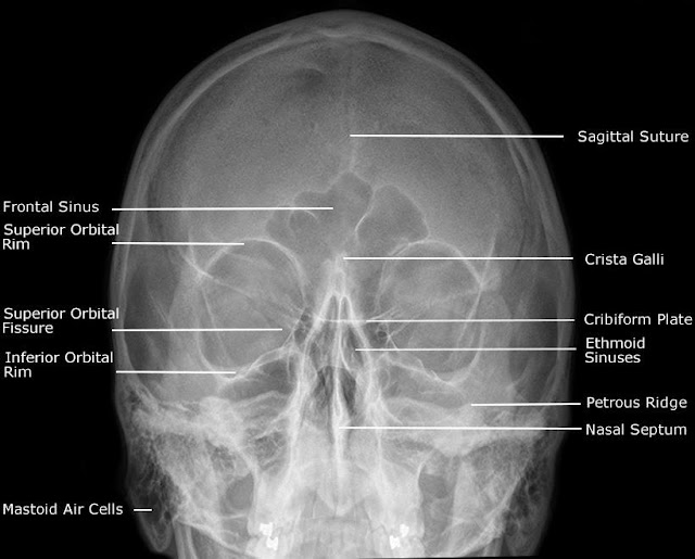
Postero-Anterior Caldwell View
Occipito Mental (OM) (Waters) View
Postero-Anterior 30° (Modified Parietocanthial)
Occipito Mental 30° (OM30) View
Submentovertex (SMV) / Slit Basal / Jughandles View
Adult Facial Bones - Lateral View
Slit Townes View


Abnormalities of the Skull and Facial Bones
Cause
                           Radiological Features
Enlargement of Skull Vault
                Children
Hydrocephalus
Sutural  diastasis, increased convoluted
markings, “copper beaten skull
Raised intra cranial pressure
Bulging of fontanelle in infancy.
                Adults
Acromegaly
Enlarged frontal sinuses and mandible, erosion
and enlargement of sella turcica
Paget’s disease
Thickened skull vault, increased density of vault
and facial bones.
Increased Density
                Localised
Hyperostosis frontalis
Symmetrical thickening of the inner table of
skull vault, usually in women, of no significance.
Meningioma
Area of  localised sclerosis, possible enlarged
groove of feeding artery.
Fibrous displasia
Asymmetrical, affecting the facial bones maxilla
and base of skull.
               Generalised
Paget’s disease
Irregular sclerosis with thickened vault.
Secondary deposits. e.g. prostate & breast
Irregular sclerosis, thickened vault.
Lytic lesions
                Childhood
Secondary deposits, neuroblastoma, leukaemi
Variable appearances,  Sutural deposits may
mimic sutural diastasis.
Eosinic granuloma, Histiocytosis X
Transradient defect with bevelled edges.
                 Adults
Myelomatosis
Rounded translucent (2-10mm) multiple ‘holes’
Secondary deposits
Generally ill defined translucent patches.
Hyperparathyroidism
Mottled appearance of ‘pepper pot’ skull.
Paget’s
Sharply defined zones affecting large areas of
the vault.


Radiographic Anatomy of Mandible

Mandible - Postero-Anterior View
Mandible - Oblique View
Mandible – Lateral View
Orthopantogram (OPG)
DOWNLOAD an Article on Skull Radiographic Anatomy

Sunday, October 9, 2011

Periapical Lesions Description In Intraoral And Extraoral Radiographs.......... with pdf note for download

Periapical Lesions Description
Well-defined radiolucent lesion at the apex of the maxillary lateral incisor.  The circumscribed radiopaque (sclerotic) border signifies that the lesion is self-contained, enlarges by expansion, and is slow-growing.  There is no change in the surrounding bone.
Well-defined radiolucent lesion at the apex of the mandibular left second premolar.  The well-defined border signifies that the lesion is self-contained, and enlarges by expansion.The surrounding bone shows slight sclerosis (osteoblastic activity) which signifies resistance to the pathologic process.


Download the Note on Periapical Description

Tuesday, September 20, 2011

Anatomical Landmarks Of Panoramic Radiographs......with ppt lecture note for download

Panoramic Anatomy
                        Panoramic Anatomy Key
1.     maxillary sinus
2.     pterygomaxillary fissure                 
3.     pterygoid plates
4.     hamulus
5.     zygomatic arch
6.     articular eminence
7.     zygomaticotemporal suture
8.     zygomatic process
9.     external auditory meatus
10.            mastoid process
11.            middle cranial fossa
12.            lateral border of the orbit
13.            infraorbital ridge
14.            infraorbital foramen
15.            infraorbital canal
16.            nasal fossa
17.            nasal septum
18.            anterior nasal spine
19.            inferior concha
20.            incisive foramen
21.            hard palate
22.            maxillary tuberosity
23.            condyle
24.   coronoid process 
25. sigmoid notch
26. medial sigmoid depression
27. styloid process
28. cervical vertebrae
29. external oblique ridge
30. mandibular canal
31. mandibular foramen
32. lingula
33. mental foramen
34. submandibular gland fossa
35. internal oblique ridge
36. mental fossa
37. mental ridges
38. genial tubercles
39. hyoid bone
40. tongue
41. soft palate
42. uvula
43. posterior pharyngeal wall
44. ear lobe
45. glossopharyngeal air space
46. nasopharyngeal air space
47. palatoglossal air space


Saturday, September 17, 2011

A Note on Intraoral Radoigraphic Anatomy......with ppt download

Intraoral Radoigraphic Anatomy

The following slides identify the anatomical structures that may be seen on intraoral films. These structures are more likely to be seen when using the bisecting angle technique because of the increased vertical angulation (increased positive in the maxilla and increased negative in the mandible) commonly used with this technique. Since some of the structures may be confused with pathology, it is important to understand their normal appearance in order to make a proper diagnosis.the slides will be easier to view.

Radiographic diagnosis of Caries.....ppt

Radiographic diagnosis of Caries


Thursday, August 4, 2011

IMAGING PROTOCOLS For Evaluating Orofacial Pain

In deciding whether to use imaging during the assessment of a patient with orofacial pain, the clinician must first obtain enough information from the history and clinical examination to determine the nature and probable cause of the problem and to decide whether imaging will provide any benefits in the diagnosis and management of the patient. In many cases, it may be necessary to rule out the teeth as a source of the pain. Select intraoral and/or panoramic radiography combined with the clinical examination can generally help in this situation.
If the patient’s symptoms are suggestive of temporomandibular disorder (TMD), a thorough clinical examination may provide enough information to establish a diagnosis and to select a management strategy without imaging, even though it has been shown that clinical examination alone will not detect all cases of internal derangement.
When there appears to be a bony component to the temporomandibular problem or if the patient is refractory to conservative treatment, it may be useful to obtain information on the condition of the osseous structures of the joints. A number of techniques can be used to confirm or rule out a variety of developmental, inflammatory, degenerative, traumatic, or neoplastic processes. Panoramic radiography provides a good overview of both joints as well as the rest of the maxillofacial complex. Although only gross structural abnormalities will be visualized with this type of radiography, this degree of detail may be adequate in many cases to determine the presence or absence of bony changes. If more detail is necessary to make the diagnosis or prognosis, conventional tomography should be performed, generally in oblique sagittal views, corrected for condylar angle, in both open- and closed-mouth positions.
Coronal or frontal tomography complements the lateral view by providing images at 90˚ to the first view. While some studies show that TMJ tomography provides additional information not anticipated clinically, others show mixed results as to the effect of the findings on the management of the patient.
If an internal derangement is suspected and if patient management depends on confirmation or rejection of this diagnosis, the position and function of the articular disk can be determined by either MRI or arthrography. In most institutions, MRI is the preferred examination because it is noninvasive and can provide information about the disk as well as other soft-tissue and bony structures.
There are other causes besides TMD for pain in the head region. Panoramic radiography may be helpful in the initial evaluation of the maxillary sinus if that structure is thought to be the origin of the facial pain. The floor of the sinus is well visualized, and discontinuity of the bony margins, thickening of the mucous membrane, partial or total opacification of the antrum, and the presence of mucous retention cysts can be noted on the resultant radiograph. A full imaging evaluation of the paranasal sinuses usually requires CT although a series of plain films may be made at some institutions.
 A, Periapical radiograph of a 29-year-old woman who presented with a throbbing toothache in the maxillary left. Endodontic treatment on the first molar had been completed 10 years earlier. The second premolar was extracted due to similar symptoms 1 year before. There is a thickening of the mucous membrane above the molars and a general cloudiness of the maxillary sinus. B, Panoramic radiograph taken the same day. The unilateral clouding of the sinus is more obvious. The patient was treated with antibiotics and antihistamines, with complete resolution of symptoms within 24 hours. The round radiopaque lesion in the mandible was not investigated at the initial appointment, and the patient did not return for further treatment.


If a central lesion is suspected of being the cause of the pain, an evaluation of the skull by CT or MRI is in order. The choice of the specific imaging examination depends on the presumptive diagnosis and should be determined in conjunction with the treating clinician and the radiologist.

Friday, July 29, 2011

10 Questions and Answers About Dental Radiographic Interpretation

01.  What is the earliest radiographic sign of periapical disease of pulpal origin?
The earliest radiographic sign is widening of the periodontal ligament space around the apex of the tooth.
02 . What is the second most common radiographic sign of periapical disease of pulpal origin?
The second most common radiographic sign is loss of the lamina aura around the apex of the tooth.
03.    Describe the radiographic differences that allow one to distinguish among periapical abscess, granuloma, radicular (periapical) cyst, and an apical surgical scar.
One cannot distinguish among periapical abscess, granuloma, or radicular (periapical) cyst on radiographic grounds alone. All of these lesions are radiolucent with well-defined borders. Whereas an abscess may be expected to be less well corticated than a radicular cyst, this feature is not marked or constant enough to be of real utility. An apical surgical scar may be radiographically distinguishable from the other three lesions if there is radiographic evidence of surgery, such as a retrograde amalgam. Of course, a history should elicit the fact of surgery.
04.    How does the radiographic appearance of pulpal pathology that has extended to in volve the bone differ in primary posterior teeth from the picture commonly seen in perma nent posterior teeth?
In permanent teeth, widening of the periodontal ligament space is seen around the apex of the tooth. In primary teeth, by contrast, the infection presents as widening of the periodontal ligament space or an area of lucency in the furcation area.


05. Does any radiographic sign permit the diagnosis of a nonvital tooth?


It is frequently stated that tooth vitality cannot be determined by radiographs alone, but this is not so. The presence of a root canal filling in a tooth provides virtually conclusive proof of its nonvitality, as does the presence of a retrograde filling, usually amalgam.
06.  At times it may be difficult to distinguish between hypercementosis and condensing or sclerosing osteitis around the apex of a tooth. What radiographic feature permits a definitive diagnosis when one is confronted with this dilemma?
If hypercementosis is present, the periodontal ligament space is visible arou nd the added cementum; that is, the cementum is contained within and is surrounded by the periodontal ligament space. Condensing osteitis, by contrast, is situated outside the periodontal ligament space.
07.  What is the radiographic sign of an ankylosed tooth?
The radiographic sign of an ankylosed tooth is loss of the periodontal ligament space and lamina aura.
08.  What is the earliest radiographic sign of periodontal disease?
The earliest radiographic sign of periodontal disease is loss of density of the crestal cortex, which is best seen in the posterior regions. In the anterior part of the mouth, the alveolar crests lose their pointed appearance and become blunted. In the posterior areas, the alveolar crests usually meet the lamina aura at right angles. In the presence of periodontal disease, these angles become rounded.
09.    What is the earliest radiographic sign of furcation involvement due to periodontal disease? 
In periodontal disease, one may see the loss of a cortical plate, either the buccal or lingual plate, on an intraoral film. The plate may be lost so that the crest now occupies a position apical to the furcation. This appearance, however, does not permit a diagnosis of furcation involvement. Widening of the periodontal ligament space in the furcation area is the earliest radiographic sign of furcation involvement.
10 . What is the radiographic differential diagnosis of a radiolucency on the root of a peri odontally healthy tooth?
Internal resorption, external resorption, and superimposition are the most common causes. Note that the question refers to a periodontally healthy tooth. If bone loss has resulted in exposure of the root, caries and abrasion, among other potential possibilities, enter the picture.

Share

+1 this blog

you might also like

Related Posts Plugin for WordPress, Blogger...