Bone Swellings

Bone swellings are lesions that characteristically
present as asymptomatic hard lumps, covered by normal epithelium. Developmental
disorders, benign and malignant tumors are included in this group of lesions.
- Torus mandibularis
- Torus palatinus
- Multiple exostoses
- Osteoma
- Osteosarcoma
- Chondrosarcoma
- Burkitt lymphoma
- Multiple myeloma
- Paget disease
- Odontogenic tumors
Torus Mandibularis
Definition and etiology Torus mandibularis is a
developmental malformation of unknown etiology.
Clinical features It presents as an asymptomatic
bony swelling, covered by normal mucosa. The lesion displays slow growth during
the second and third decades of life. Characteristically, the lesions appear bilaterally
on the lingual surface of the mandible, usually in the area adjacent to the
bicuspids. The diagnosis is based on clinical criteria.
 |
| Torus mandibularis |
Treatment Unnecessary unless full denture
construction is required.
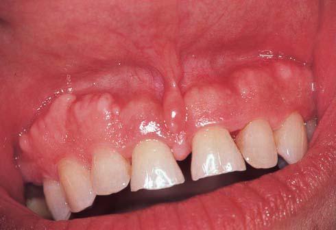
Torus
Palatinus
 |
| Torus palatinus at the midline of the hard palate |
Definition and etiology Torus palatinus is a
developmental malformation of unknown etiology.
Clinical features It presents as a slow-growing,
nodular, lobular or spindled, asymptomatic bony swelling covered by normal
mucosa. Characteristically, the lesion appears along the midline of the hard
palate.It occurs more often in women, and usually appears during the third
decade of life. The diagnosis is based on the clinical findings.
Treatment Unnecessary unless full denture construction is required.
Multiple Exostoses
Multiple exostoses may occur on the buccal surface of the
maxilla, and rarely on the mandible. Clinically, the lesions appear as multiple
asymptomatic bony swellings. The diagnosis is based on the clinical findings.
 |
| Multiple exostoses on the maxilla. |
Treatment Unnecessary unless
full denture preparation is required.
Osteoma
Definition Osteoma is a benign
neoplasm that consists of mature compact or cancellous bone.
Etiology Unknown.
Clinical features
It presents as an asymptomatic, slow-growing bony swelling
of the jaws. The size ranges from a few millimeters to several centimeters.
Multiple jaw osteomas are a common feature of Gardner syndrome.
 |
| Gardner syndrome: osteoma of the mandible. |
Laboratory tests Histopathological
examination, radiography.
Differential diagnosis Exostoses,
osteosarcoma.
Treatment Surgical excision.
Osteosarcoma
Definition Osteosarcoma is the
most common primary malignant neoplasm of bone.
Etiology Unknown.
Clinical features
The jaws are affected in 6–7% of cases, and usually during
the third decade of life. Both jaws are affected equally and it is more common
in men. Clinically, the lesion presents as a rapidly growing hard swelling that
progressively produces facial deformity. Pain, paresthesia, tooth loosening,
and nasal obstruction may also occur.
 |
| Osteosarcoma of the upper jaw, presenting as a hard swelling. |
Laboratory tests Histopathological
examination, radiography, CT scans.
Differential diagnosis Chondrosarcoma,
Ewing sarcoma, metastatic tumors, odontogenic tumors and cysts, giant-cell
tumor.
Treatment Surgical excision and
supplementary radiotherapy and chemotherapy.
Chondrosarcoma
Chondrosarcoma is more common in men than in
women, between 30 and 60 years of age. Clinically, it appears as a painless
hard swelling that progressively enlarges, causing extensive bone destruction with
pain and loosening of the teeth.
Burkitt Lymphoma
Definition Burkitt lymphoma is a
high-grade malignant B-lymphocyte lymphoma.
Etiology Epstein–Barr virus is
closely associated.
Clinical features
The malignancy is prevalent in central Africa (the endemic
form), and usually affects children 2–12 years of age. Cases have also been
observed in other countries (the nonendemic form), and recently in patients
with AIDS. The jaws are the most common site of lymphoma (60–70%). Clinically,
it presents as a rapidly growing hard swelling that causes bone destruction,
tooth loss, and facial deformity.Pain, paresthesia and large ulcerating or
nonulcerating masses may also be seen.
|
| Burkitt lymphoma, facial deformity. |
 |
| Burkitt lymphoma, gingival mass |
 |
| Burkitt lymphoma on the gingiva in a young patient with AIDS |
Laboratory tests Histopathological
examination, radiography.
Differential diagnosis Central
giant-cell granuloma, ossifying fibroma, other non-Hodgkin lymphomas, and
odontogenic tumors.
Treatment Chemotherapy,
radiotherapy.
Multiple Myeloma
Definition Multiplemyeloma is a
relatively rare malignant plasma-cell disorder.
Etiology Unknown.
Clinical features The
malignancy is more common in men over 50 years of age, and the jaws are
affected in about 30% of cases. Clinically, it presents with bone swelling,
tooth mobility, pain, and paresthesia. A painless soft swelling, usually on the
alveolar mucosa and gingiva, may develop as part of the overall disease
spectrum.
 |
| Multiple myeloma, swelling on the gingiva |
Laboratory tests Bone-marrow
biopsy, radiography, serum and urine protein electrophoresis.
Differential diagnosis Plasmacytoma,
non-Hodgkin lymphoma, Ewing sarcoma, leukemia, Langerhans cell histiocytosis.
Treatment Chemotherapy,
radiotherapy.
Paget Disease
Definition Paget disease, or
osteitis deformans, is a chronic, relatively common disorder characterized by
uncoordinated bone resorption and deposition.
Etiology Unknown.
Clinical features Clinically,
the signs and symptoms develop gradually and are characterized by bone pain,
headache, deafness, visual disorders, dizziness, and progressive bone
enlargement. Progressive expansion of the maxilla and the mandible lead to
symmetrical thickening of the alveolar ridges.
 |
| Paget disease, enlarged maxilla |
Edentulous patients may complain that their dentures do
not fit due to alveolar enlargement.
 |
| Paget disease, alveolar enlargement |
Delayed wound healing, bleeding, and osteomyelitis after
tooth extraction may occur. The maxilla is more frequently affected than the
mandible. Malesare more often affected than females. Two major forms of the
disease are recognized: (a) the monostotic, and (b) the polyostotic.
The clinical diagnosis should be confirmed by a histopathological and radiographic
examination. Elevations of serum alkaline phosphatase and urinary
hydroxyproline levels are common findings.
Differential diagnosis Fibrous
dysplasia, osteosarcoma, multiple exostoses, fibro-osseous lesions.
Treatment Most cases require no
treatment. Calcitonin and bisphosphonates may slow the pathological process.
Odontogenic Tumors
Definition Odontogenic tumors
are a group of lesions that originate from odontogenic epithelium and
ectomesenchyme.
Etiology Unknown. Some are
neoplasms and others hamartomas.
Classification On
the basis of the tissue of origin, three major varieties are recognized: (a) tumors
of odontogenic epithelium, (b) tumors of odontogenic ectomesenchyme,
and (c) mixedod ontogenic tumors.
Clinical features Most
odontogenic tumors are usually asymptomatic for long time and are discovered
only during a routine radiographic examination. However, with time they may
form a usually painless slow-growing swelling or expansion of the mandible or
the maxilla.
 |
| Odontogenic myxoma, expansion of the retromolar area |
 |
| Extraosseous calcifying epithelial odontogenic tumor presenting as a gingival mass |
The clinical signs and symptoms are not diagnostic and
the final diagnosis should be made by radiographic and histopathological examinations.
Differential diagnosis Different
varieties of odontogenic tumors, odontogenic cysts, osteosarcomas,
chondrosarcomas, multiplemyeloma.
Treatment Surgical excision.


