
Showing posts with label dental anatomy. Show all posts
Showing posts with label dental anatomy. Show all posts
Thursday, September 29, 2011
Saturday, September 24, 2011
A Note on Dental Anatomy of Premolars
General Features of Premolars


Dental Anatomy Lecture Note on Premolars
— They have at least two cusps.
§ one large buccal cusp,
§ Smaller lingual cusp
— The lower second premolar may- sometimes- have two lingual cusps.
Maxillary 1st Premolar


Maxillary 2nd Premolar


Mandibular 1st Premolar

Mandibular 2nd Premolar

Saturday, September 10, 2011
HEAD AND NECK DEVELOPMENT, PHARYNGEAL ARCH, GROOVE, AND POUCH DEVELOPMENT
In the human embryo, the stomodeum or primitive mouth cavity, a shallow depression lined with ectoderm, is separated from the cephalic end of the pharyngeal gut by the buccopharyngeal membrane, which ruptures during the fourth week of gestation.

Frontal view of an embryo at 4 to 5 weeks of age. Observe the branchial arch formation and the ruptured buccopharyngeal membrane.
Just anterior to the buccopharyngeal membrane, a midline diverticulum known as the Rathke pouch develops in the oral ectoderm of the roof of the primitive oral cavity. This evaginating pouch comes in contact with a pouch developing from the floor of the diencephalon. Further development of these two opposed structures gives rise to the pituitary gland.
During the fourth and fifth weeks of gestation, as the buccopharyngeal membrane is degenerating, communication is established between the future oral cavity and the pharynx. Concomitant with this event, out-pouchings of the pharyngeal gut develop just posterior to the ruptured buccopharyngeal membrane, forming five pairs of pharyngeal pouches that invade the mesenchyme laterally. Although it was once thought that the neural crest cells of the pharyngeal arches were responsible for patterning skeletal components of the arches, it is now clear that the endodermal lining of the pharyngeal pouches regulates this patterning.
Concurrent with the formation of the pouches in the pharyngeal wall, four pairs of grooves develop around the stomodeal neck area on the lateral surface of the embryo. These pharyngeal grooves (branchial grooves) invade the underlying mesenchyme approximating, but not contacting, their corresponding pharyngeal pouches. Invasion by the pouches and the grooves produces a condensation of mesenchymal tissue interposed between successive pairs of pouches and grooves (clefts). These five bars of condensed mesenchymal tissue are the pharyngeal (branchial) arches (see Figs. 5-2, 5-3A, 5-3B, 5-3C, and 5-4).

Developing face. (A) Fourth week. (B) Fourth to fifth week. (C) Fifth to sixth week. (D) Sixth to seventh week. Note how the nose develops and the eyes appear more anteriorly placed, illustrating the nasolacrimal groove.

Scanning electron micrograph of a stage 15 (8.0-mm) human embryo in frontal view. Observe the maxillary processes, mandibular process, hyoid and third pharyngeal arch processes, and nasal pits. ×52.

Scanning electron micrograph of a stage 17 (11.7-mm) human embryo in lateral view. Observe the nasal pits, median and lateral nasal processes, maxillary processes, mandibular process, and hyoid arch. ×57.
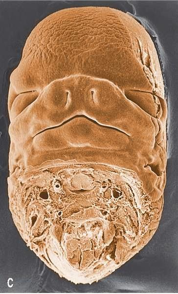
Scanning electron micrograph of the face of a stage 18 (17.5-mm) human embryo in frontal view. Observe the fused processes making up the nose, the maxillary processes, and the mandibular processes delineating the mouth. ×14.

Early development of the pharyngeal grooves and pouches. (A) Early development. (B) Later development. Observe the second arch overgrowing the second, third, and fourth grooves leaving a cervical cyst in drawing B. Note the diverticula of pouches three and four as each develops dorsal and ventral prolongations.
The mesenchymal cells forming much of the ventral regions of the developing pharyngeal arches are derived from neural crest cells originating from the vicinity of the midbrain region and the rhombomeres. During early pharyngeal arch formation, these cells express homeobox gene (Hox gene) products indicative of the region of their origin.
Continued development and growth of the arches results in their fusion in the anterior midline. Each arch will develop its own bone, cartilage, vascular, muscular, and nerve components. The arches form sequentially from rostral to caudal, with the first arch being most highly developed and the last arch being poorly developed. The first and second arches are named “mandibular” and “hyoid” arches, respectively, whereas the remaining three are unnamed. The discussion of pharyngeal arch derivatives that follows is summarized in Table.
Pharyngeal Arch Derivatives and Their Innervation | |||||||||||||||||||||||||||||||||||
| |||||||||||||||||||||||||||||||||||
a The origin of some of the muscles of the pharynx is as yet unclear. The pharyngeal constrictors may in fact receive innervation from more than one source.
b Although this named branch of the vagus nerve is traditionally described as the motor innervation to the identified laryngeal muscles, it is important to remember that the motor fibers of the vagus nerve, at least in this region, are actually motor fibers contributed to the vagus nerve from the cranial portion of the accessory nerve (cranial nerve XI).
c Several of the pharyngeal and associated muscles of the soft palate are innervated by branches of the pharyngeal plexus—a complex of the nerves located on the posterior aspects of the pharynx consisting of contributions from cranial nerves IX and X (IX is sensory, whereas X is motor, although these motor fibers are contributed from XI). Fibers from the superior cervical gaglion also contribute to the pharyngeal plexus, serving vasomotor functions.
Thursday, August 25, 2011
A Note On Tooth Development(Odontogenesis)......With Diagrams and Interactive Videos
Odontogenesis


Tooth Devolopment 3D video to understand tooth development
Tooth Development Video Explanation using Diagrams
Histology Of Tooth Development Video
Tooth formation is highly regulated at the molecular level
• terminal differentiation of specific cell types
• epithelial-mesenchymal interactions
• secretion of specific extracellular matrices
• controlled processing of those matrices
• regulation of ion deposition
• mineralization of the dental tissues
Tooth Formation is dependent on
Genetic Factors
Hundreds to several thousand genes likely involved (polygenic)

Tooth development is represented by four stages: bell stage (A), initial protein-matrix secretion (B), regression of dental epithelium (D), and tooth eruption (E). Sagittal sections were stained with alcian blue, hematoxylin, and eosin. Arrowheads, dental epithelium. Asterisks, artificial gaps. (Scale bars: 10 μm.) ISH analysis revealed the expression of SCPP1 (G),SCPP2 (H), SCPP3A (I), SCPP3B (J), SCPP4 (K), SCPP5 (L), COL1 (M), and SPARC(N) in various developmental stages. The expression of SCPP genes is summarized for the initial secretory stage (C) and for the eruption stage (F). IDE, inner dental epithelium; IPE, inner pharyngeal epithelium; Ms, mesenchyme expressing COL1 and SPARC; Od, odontoblasts; ODE, outer dental epithelium; OPE, outer pharyngeal epithelium; PRM, protein matrix.
Environmental Factors
Nutrition
Physical phenomenon
Infection
Molecular Determinants of Tooth Formation
• Over 10,000 genes involved in making a tooth
• Most genes involved in odontogenesis are expressed in non-dental tissues
• Some genes are relatively specific for development of the dental tissues (e.g. amelogenin gene)
Environmental Influences of Environmental Influences of Amelogenesis
• Nutrition
Major and minor components
• Calcium, phosphorus, protein, fluoride etc…
• Hypoxia
• Hyperthermia
• Infection
Congenital rubella, syphillus, CMV, etc…
• Physical Determinants
Space
Trauma
Interactions Leading to Developmental Dental Defects
Stages Required for Tooth Formation
• Initiation
• Histodifferentiation
• Morphodifferentiation
• Apposition
Secretory Phase
Transition Phase
Maturation Phase
• Stages are not discrete for any given tooth.
• Teeth develop over years beginning with the coronal portion of the crown.
• Can be mineralizing at the cusp tips while cervically cells are differentiating.

Tooth Development Video Explanation using Diagrams
Histology Of Tooth Development Video
Subscribe to:
Posts (Atom)
