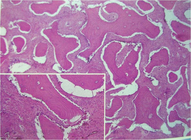
Fibro-osseous lesions
• Diverse group of processes characterized by replacement of normal bone by fibrous tissue containing newly formed mineralized product. (Not a specific diagnosis)
• Includes lesions of,
– Developmental
– Reactive
– Dysplastic
– Neoplastic
CLASSIFICATION
– Fibrous dysplasia
– Cemento-osseous dysplasia
• Focal
• Periapical
• Florid
– Ossifying fibroma
Fibrous dysplasia
• Defined as a non-neoplastic, primary disorder of bone in which normal medullary bone is replaced by a variable amount of structurally weak fibrous and osseous tissue.
• A developmental tumor like condition that is characterized by replacement of normal bone by an excessive proliferation of cellular fibrous connective tissue intermixed with irregular bony trabeculae.
• Mutation involving GNAS1 gene
Clinical features
• FD can manifest as,
– Involve only one bone (Monostotic) – mutation in post natal life (confined to one site)
– Involve multiple bones (Polyostotic) – mutation of skeletal progenitor cells and their progeny will involve development of multiple bones.
– Multiple bone lesions in conjunction with cutaneous and endocrine abnormalities – mutation of undifferentiated stem cells in early embryonic life (osteoblasts, melanocytes and endocrine cells)
Monostotic FD
• Limited to a single bone and can stabilize by puberty.
• 80-85% of cases – jaws more commonly affected - maxilla
• Diagnosed during 2nd decade
• Males and females are equally affected
• Painless, slow growing swelling of affected area
• Teeth are displaced but remain firm.
• Mandibular – truly monostotic
• Maxilla – can involve adjacent bones – zygoma, occipital, sphenoid – Craniofacial FD
• Ground glass opacification – poorly calcified bone trabeculae arranged in a disorganized pattern.
• Margins blend into the normal bone
• Expansion of buccal, lingual plates with bulging of lower border
• Superior displacement of inferior alveolar canal
• Obliteration of maxillary sinus
• Increased density in base of skull – occiput, sphenoid, roof of orbit and frontal bones – FD of skull.
Polyostotic FD
• Involvement of 2 or more bones and continues to grow.
• Jaffe-lichtenstein syndrome – polyostotic FD with café au lait (Coffee with milk) pigmentation.
• Mccune-Albright syndrome - polyostotic FD with café au lait pigmentation and multiple endocrinopathies – sexual precocity in females, pituitary adenoma or hyperthyroidism.
Clinical features
• Facial asymmetry
• Symptoms are related to long bone lesions like pathologic #
• Involvement of upper portion of femur leads to leg length discrpancy – Hockey stick deformity
• Café au lait pigmentation – well defined, unilateral, tan macules on trunk and thighs. The margins are irregular whereas in NF, the margins are smooth.
histopathology
• Irregularly shaped trabeculae of immature (woven) bone in a cellular, loosely arranged fibrous stroma.
• Trabeculae are not connected with each other, assume curvilinear shapes – Chinese script writing
• Arise by metaplasia and are not surrounded by osteoblasts.
• Monotonous pattern throughout the lesion.
• Fuses directly with the normal bone without any line of demarcation.
• Can undergo progressive maturation.
Treatment
• Surgical resection
• Disease tends to stabilize and stops enlarging when skeletal maturation is reached.
• Regrowth of the lesion in 25-50% of cases – more in younger pts


After remove FD, what next treatment for the patient? What rehabilitation? Tq
ReplyDeleteI've been living with Parkinson’s for several years. When my usual medications started losing their effectiveness, I decided to explore alternative options. I came across a natural approach from NaturePath Herbal Clinic. Honestly, I was skeptical at first — but after about four months on their herbal program, I started to notice meaningful changes: reduced tremors, less muscle stiffness, and better sleep quality.
ReplyDeleteIt’s not a cure, but for me, it’s been a noticeable improvement in daily life. If you're considering natural or complementary options alongside traditional treatment, this might be something worth checking out: www.naturepathherbalclinic.com.